The Radiology Department provides comprehensive outpatient radiologic imaging services.
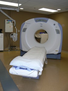
 JMHS is proud to offer a 64 slice GE scanner. This allows for more imaging shorter scan times and shorter breath holds for patients.
What is a CT Scanner?
A CT scan is a specific kind of X-ray. The scanner makes cross-sectional images of body tissues.
How is the test done?
You may wear your own clothes for the scan as long as there are no metal objects (such as zippers or snaps) near the body part to be scanned. Sometimes a contrast media is used to give more information about your body. If contrast is needed, a nurse will start an IV in a vein, usually in the arm or hand. This involves a small poke using a small IV needle. Once the IV is in place, the needle is removed and a tiny plastic tube stays in the vein during the scan. You will see the large scanning machine with an opening in the middle. You will lie on a padded imaging bed, which moves in and out of the opening smoothly.
The room lights may be dimmed during the scan. It contrast is used; it will be given into the IV during the scan using a power injector. You may experience a warm sensation, a metallic taste in your mouth, or a feeling of needing to urinate as the contrast is given. This is perfectly normal and happens most of the time. The scanner will not touch you. It makes a whirring noise as it scans.
You must hold very still and not talk during the scan. You might be asked to hold your breath, we ask you that you do the best you can and breathe when the machine tells you to.
What should I do before the scan?
You should have nothing to eat or drink at least 4 hours before the scan. If oral contrast is needed, further instruction will be provided by the nurse or x-ray technologist.
What to expect after the test?
After the scan, if IV contrast was administered it will pass through the kidneys and unnoticed in your urine. It’s important to drink extra water after the scan to help pass the contrast. The ordering doctor will contact you with results.
Approximate time of exam is 5 minutes.
Please call 320-769-4323 Ext 2194 for any questions or concerns regarding your exam.
What’s an MRI?
An MRI scanner creates three dimensional images of body tissues using a large magnet and radio waves. It does not use radiation. There are no known harmful effects from having an MRI scan. We use a Wide Bore machine that can accommodate patients up to 500lbs. The larger tube provides for a less closterphobic experience.
How is the test done?
You will go out to a mobile MRI unit that is on a truck. The magnet is always on the room. Due to the strength of the magnetic field all metal objects such as barrettes, bobby pins, jewelry and credit cards must be used to give more information about your body. If contrast is needed, an IV will be started in the arm or hand by the MRI technologist. The scanning machine is large with an opening in the middle. You will be lying on the imaging bed, which moves in and out of the opening smoothly. A coil will be placed over the part of the body being scanned. The scanner will not touch you. It makes a loud thumping or tapping sounds. You will be given earplugs to wear. You must hold very still during the test.
What to expect after the test?
If IV contrast was injected, drink plenty of water after the test to help flush it through the kidneys. The ordering doctor will contact you with results within a few days.
MRI is provided by Shared Medical Services (SMS) out of Cottage Grove, WI.
Approximate time of each scan is 45minutes. Available Tuesday Mornings.
Approximate exam time is 45 minutes. Available by appointment only.
How is the test done?
The ultrasound technologist will bring you into an exam room and you will lie on a padded table. The lights in the room may be dimmed to help the technologies see his pictures better. The technologist will apply warm gel to the transducer and on the area to be scanned. The transducer sends sound waves through the body.
As the sound waves travel through the body, they send echoes back through the transducer to the computer. The computer makes images of the sound wave patterns. Ultrasounds are usually painless; you may feel slight pressure as the transducer is moved over the skin.
What to do before the test?
Depending on the test ordered you may be asked to not eat or drink anything including water before the test. A nurse will provide you with those instructions prior to the exam date.
After the test?
You may go back to normal eating and activity after an ultrasound. You will receive the results from the ordering physician.
Ultrasound is available Monday through Friday.
Types of Ultrasounds we offer
- Abdominal
- OB/GYN – 3D & 4D OB ultrasound
- Breast
- Pediatric
- Vascular
Each exam takes approximately 30 minutes.
 A mammogram is a look for changes in the breast that are not normal by use of a low-dose x-ray exam. A doctor called a radiologist to will examine the results and be able to see changes in breast tissue that cannot be felt during a breast exam. All women should have their breast examined. If symptoms appear, such as a change in the shape or size of a breast, a lump, nipple discharge, or pain, you should see your physician. Johnson Memorial Health Services is pleased to provide digital 2D and 3D mammograms in-house.
3D Mammography
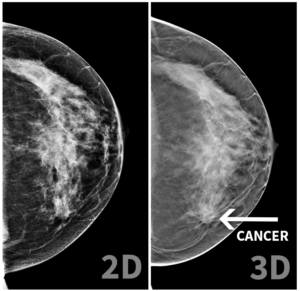
 3D mammography is a digital exam that produces a 3D image of the breast. It gives us a “stack” of images in layers to better evaluate the breast tissue. The 3D exam also generates a standard 2D digital mammogram for screening exams. 3D mammography allows breast radiologists (doctors with training in breast imaging) to examine breast tissue more thoroughly. Fine details are more clearly visible, no longer hidden by the tissue above and below. 3D images give a better view of the size, shape, and location of abnormalities. This results in a better exam. Studies of 3D mammograms have shown better detection of breast cancer in nearly all breast densities. It also decreases the number of patients who need to return to have more images taken. The radiation dose is similar to that of a standard digital 2D mammogram.
Will this cost me anything?
There is an additional JMHS facility charge of $55 for 3D mammography, and an additional $86 for Midwest Radiology for the reading. Many insurances cover this cost as part of preventative screening benefit, however some do not. There may be an additional co-pay and/or you may see this amount applied to your deductible. You will want to consult your insurance carrier about your specific plan benefits to verify your coverage for 3D mammography prior to your exam. The number can be found on the back of your insurance card. Take a few minutes now to call your insurance carrier to find out if 3D imaging is covered for you. If you’re told an order is needed or you can have it only if it’s medically necessary, please tell this to your technologist.
All mammograms are interpreted by Midwest Radiology with reports sent to your practitioner.
Contact Radiology at 320-769-4323 to schedule an appointment.
Learn more about MammogramsWhat is a bone scan?
 A bone scan studies the bones in your body and detects fractures, infections or other bone problems. The test gives very radiation. A radioisotope is given into your vein. You will not feel the isotope or have any side effects from it. The camera detects gamma rays (invisible radiation) coming from the radioisotope ad creates the images on the computer.
How is the test done?
The technologist will administer the radioisotope into your vein. Depending on the test your doctor has ordered, a first set of images may be taken right after the radioisotope is injected. This may take about 15 minutes. Whether or not the first images are taken, you will be asked to return in 3 hours to complete the exam. You may leave, eat and drink before and after the isotope is given.
When image are taken, you will lie on the imaging bed with the camera very close to your body. These sets of images will take 25 to 55 minutes.
What to expect after the test?
You can usually go back to routine activities unless told otherwise. Your ordering doctor will contact you with results within a few days.
Service is provided by Avera in Sioux Falls, SD.
Approximate exam times vary. Available by appointment only.
The scan is performed by trained technologists and takes approximately 10 minutes to complete.
